Tying up
Tying-Up – what we’re talking about?
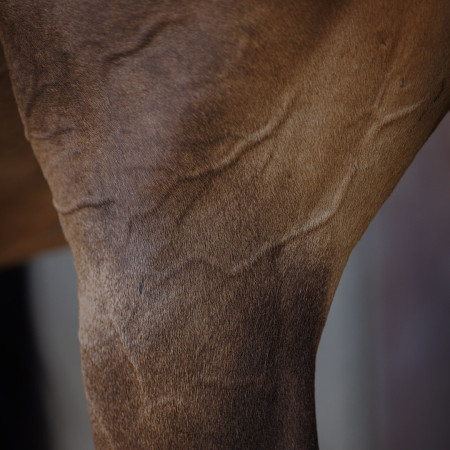
Tying-up (also called Azoturia or exertional rhabdomyolysis) is a syndrome that describes the occurrence of abnormal muscle stiffness due to muscle damage following intensive exercise. Basically, tying-up refers to severe muscle cramping – primarily of the large muscles of the hind limb and the back – leading to a buildup of metabolic waste which cannot be removed rapidly enough and therefore cause changes in acid-base balance, energy metabolism, electrolyte balance while resulting in muscle fatigue, abnormal contracture, and even muscle cell damage. This can be extremely painful for affected horses who can react colicky beside heavy sweating, increased heart and breathing rate, unwillingness to move, brown colored urine.
Tying-up usually affects high performance horses, rested for two or more days but still fed a diet rich in carbohydrates and low in fat. Typically clinical findings mostly occur after the first 20 minutes of intensive work. Tying-up also can be triggered by suddenly increased work intensity of untrained horses or even by abrupt dietary changes or imbalances in performance horses.
Tying-up is one of the most common muscle disorders in horses.
What’s the reason for tying-up in horses?
Overall, tying-up can be caused by a variety of factors and apparent environmental stimuli can trigger this syndrome. Most affected horses, especially by repeated muscle problems, are those fed on a diet rich in starch even if they pause from their usual training intensity. This may be related due to interaction with the storage of glycogen within the muscle tissue.
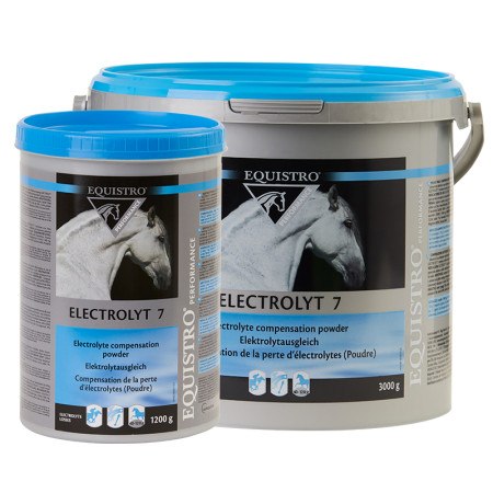
Furthermore, electrolyte imbalances due to dietary inadequacies or heavy sweating during exercise are suspicious to cause tying-up as well (Thoroughbreds are very susceptible to).
Overexcitement also can induce episodes of tying-up, which affects young fillies the most. To prevent such horses form the occurrence of further episodes, daily exercise and a stressless environment beside a well-balanced mineral and vitamin supplement combined with a minimum of carbohydrates and high-quality hay should be granted.
A specific cause of tying-up is PSSM (polysaccharide storage myopathy). It is characterized by an abnormal accumulation of glycogen molecules (storage form of glucose) within the skeletal muscle cells. PSSM can be categorized into two forms: PSSM type I and PSSM type II. Horses with type I PSSM are suffering from a genetic mutation of the GYS1 gene expressing an enzyme that excessively converts glucose into glycogen which is stored in the muscle cells, rendering the glucose molecules unavailable to meet the energy needs of the muscle. Type 1 PSSM occurs in many Quarter Horses, Paint Horses, Appaloosa Horses, warmblood breeds and many pony breeds. Affected horses develop muscle stiffness / cramping and soreness during light exercise due to an energy deficit in the muscles. Type 2 PSSM is assumed to refer to a genetic cause as well which still is unknown yet. It has a similar microscopic appearance than type 1 PSSM and occurs in Quarter Horse-related breeds, Warmbloods and Thoroughbreds. While type 1 PSSM can be diagnosed by a DNA-based test from blood or hair, type 2 PSSM requires a muscle biopsy. Treatment of both PSSM forms includes providing horses with an alternative source of energy (e.g., fat from oil) instead of carbohydrates for stabilizing blood sugar levels and keep up the energy metabolism of the muscles. Horses with mild clinical findings can return to their full performance potential with dietary and management changes, including continuous daily exercise without extended periods of inactivity. Horses with PSSM have at least a 50 percent chance to pass the genetic mutation on to the offspring.
What to do when horses are tying-up?
Horses with tying-up can show a variety of symptoms of mild to moderate or even severe intense which typically occur after 15-20 minutes of exercise or after finished exercise. Occurring symptoms indicating an episode of tying-up are:
- Muscle cramping (muscles of the back and hind limb are hard and stiff)
- Shortened, stiff gait or even unwillingness to move (severe cases)
- Excessive sweating
- Increased heart and breathing rate
- Pain face (raised nostrils, ears set back, tense cheek muscles) due to muscle pain
- Pain-induced colic-like symptoms (flank watching / kicking, poor appetite, change in drinking behavior, stretching out)
- Posturing to urinate frequently (brown-colored urine in severe cases due to the destruction of muscle cells and the release of Myoglobin)
If a horse is suffering from tying-up, the following measures should be undertaken:
- Stop exercising immediately and take the horse to the stall but do not force the horse to move if it refuses walking
- As tying-up is considered as veterinary emergency, so a vet should always be consulted when episodes are occurring
- Rub the horse dry (in case of heavy sweating), then blanket the horse if temperatures are low
- Rehydrate the horse (electrolytes + free access to water) if dehydration due to heavy sweating need to be assumed
- Remove feed, especially grain, from the stall (only hay should be provided until the symptoms occur)
For diagnosing tying-up, the vet depends on blood work, urinalysis and a muscle biopsy in some specific cases. An increased concentration of the muscle specific enzymes CK (ceratine kinase), AST (aspartate transaminase) and LDH (lactate dehydrogenase) which are normally located in muscle cells in combination with the occurrence of specific clinical findings is a reliable reference for the diagnosis of tying-up. The suspicion can be completed by the detection of myoglobin in the urine. The extensively increase in muscle enzymes in the blood as well as the content of myoglobin in the urine is caused by an increased release of these substances from inside muscle cells due to their enormous damage.
Long-term management of tying-up horses
Medical treatment of tying-up depends on the severity of the symptoms and aims to reduce pain (NSAIDs), inhibit further muscle damage and protect the kidneys.
The training program should be designed according to the individual condition of each horse. Training intensity and duration always should be increased slowly and steady to avoid overexertion. As tying-up can be triggered by stress (e.g., transport, separation from other horses), the horse’s environment should be kept as comfortable as possible. A well-balanced diet low in non-structural carbohydrates (10-15% of starch maximum) is important and can also prevent electrolyte imbalances and vitamin deficiencies. Electrolytes are essential for normal muscle function and must additionally be supplemented if horses are sweating excessively after exercise. Adding vitamin E and other antioxidants to the diet of horses predisposed to tying-up can protect the muscle membrane. The energy density of the diet can be increased by supplementing fat (oil) as well as highly digestible fiber like beet pulp for instance.
Dr. med. vet. Julie Dauvillier
Dr. med. vet. Caroline Fritz