Navicular Syndrome
Though the term “navicular disease” is commonly used, the exact term is « podotrochlear syndrome » which refers to foot pain located in the heels and caused by a complex of pathologic mechanisms.
Podotrochlear syndrome - what we're talking about?
The navicular bone is beside the bursae, the deep flexor tendon and some surroning ligaments part of the so-called podotrochlea structure. Although, the navicular bone may be the source of the pain, the surrounding structures (tendons, ligaments, bursae) are also discussed to have a main influence on this pathology as all of these structures make up the podotrochlear apparatus and can be the source of lameness.
The word “syndrome” is used because, even if symptoms are similar, there are multiple causes of the navicular syndrome, which are usually unknown.
Predisposing factors are well-known
- Horse breed (sport horses are mainly affected)
- Foot conformation (long toe and elusive heels)
- Overfeeding during growth, excessive work at the end of growth
- Aggravating factors (e.g., training on irregular or hard ground, repetition of heavy loads, inadequate trimming/shoeing)
Clinical findings
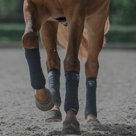
Horses with navicular bone pain usually show intermittent lameness. It is more noticeable when walking on hard ground and potentially affects both forelimbs. Lameness is usually worse at the beginning of training and the day after strenuous activity. Owners may notice a decline in performance, a horse taking shorter strides, or a horse which does not want to make sharp turns.
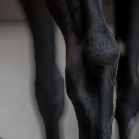
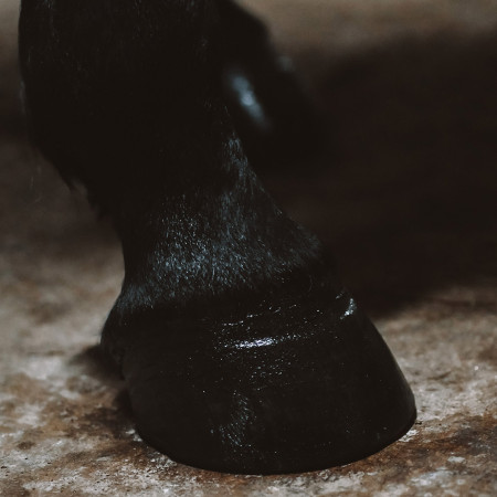
At rest, when a single limb is affected, the horse often holds the affected limb out in front of him to relieve pain from the navicular area. This posture gradually causes waves that appears on the affected foot’s hoof and an increase in the heel’s size.
Diagnosis
A navicular syndrome diagnosis is made in two steps:
- A first orthopedic exam allows the localization of the lameness
- A second medical imaging defines its origin.
Many mechanical tests help the diagnosis of foot lameness:
Hoof tester
Pressure is applied on the affected foot with a specific clamp to localize the painful area. Test is positive if the horse withdraws his foot under the pressure. This test is often positive on the heels of a horse with navicular disease.
Board test or extension test
The horse’s foot is set on the end of a board. An operator keeps the other limb raised while a second operator lifts the board gradually to a 40° angle. The result is positive if the horse tries to get off of the board. In the majority of cases, this test is positive on a horse affected by navicular disease.
Flexion test
Distal articulations are put in flexion for 45 seconds to 1 minute and then the horse is led to trot. Flexion is positive if lameness is increased after the test. In most cases, a horse with navicular disease tests positive.
To precisely locate the lameness, anesthesia called « palmar digital analgesia » is used to localize the spot of pain. A local anesthesia of the foot is performed, and then the horse is led to trot. In the case of podotrochlear syndrome, the anesthesia eliminates the lameness in 80 to 90% of cases.
Afterwards, the diagnosis is specified with x-ray. Preparation of the foot is important to develop good quality images and the horse may have to be unshoed. Images of multiple orientations are taken in order to perfectly visualize the navicular bone and the articulation between the second and third phalanx.
Imaging diagnostic
Many abnormalities can be observed in the radiographs: the appearance of bone fragments on the ligament insertion on the navicular bone; radiolucent areas on the distal part of the bone; increase or decrease of bone density or damage to the bone surface in contact with the deep digital flexor tendon.
To make a more precise diagnosis, and especially in the case of deep digital flexor tendon affection in the foot or in the navicular bursae, ultrasound can be used.
Recently, MRI has become the gold standard, and the examination of choice. It allows a precise evaluation of soft tissues and bones in the foot.
Treatment opportunities
Treatment of the navicular syndrome depends on the cause. However, when the bone is affected, treatment is palliative and not curative. It must be adapted to each horse based on its use, on the owner’s choice and on the extent of diagnosed lesions.
Horses with severe lameness must be kept at rest for a while and must receive anti-inflammatory drugs. The work-load has to be adapted and managed: good walking warm up, work on soft and even ground, and work in straight lines with no sharp turns.
Good trimming and shoeing is an important aspect of the treatment. Horseshoes should reduce footwork (facilitate rolling, lighten the foot) and decrease injuries in the heel area. As such, the toe must be trimmed and a shoe with a good rolling must be set. Heels must be protected and, in some cases, they must be slightly heightened to decrease pressure caused by the deep digital flexor tendon on the navicular bone.
Modern treatments also include regenerative therapies and intra-articular anti-inflammatory injections.
If the horse’s work is managed and proper horseshoes are set, clinical symptoms can be resolved in half of all cases. However, prognosis is always correlated with pathology type and diagnosed lesions.
Dr. med. vet. Perinne Piat